Our Services
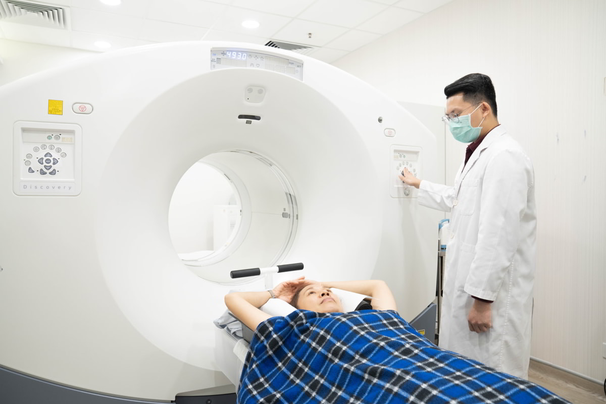
PET/CT
A PET/CT machine is a state-of-the-art imaging tool that helps physicians detect the presence and location of cancer in your body with pinpoint accuracy. The highly detailed imaging results are extremely useful for specialists to base their diagnosis on, prior to making any kind of treatment recommendation. As such, it is an invaluable tool for cancer diagnoses.
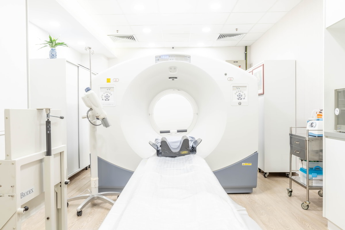
CT
A computed tomography (CT) scan is an advanced diagnostic imaging procedure that uses a combination of X-rays and computer technology to construct images of the inside of the body. This examination can provide clear images of various body structures including head, organs, blood vessels, muscles and soft tissues, all at once. The images provide thorough information to assist diagnosis.
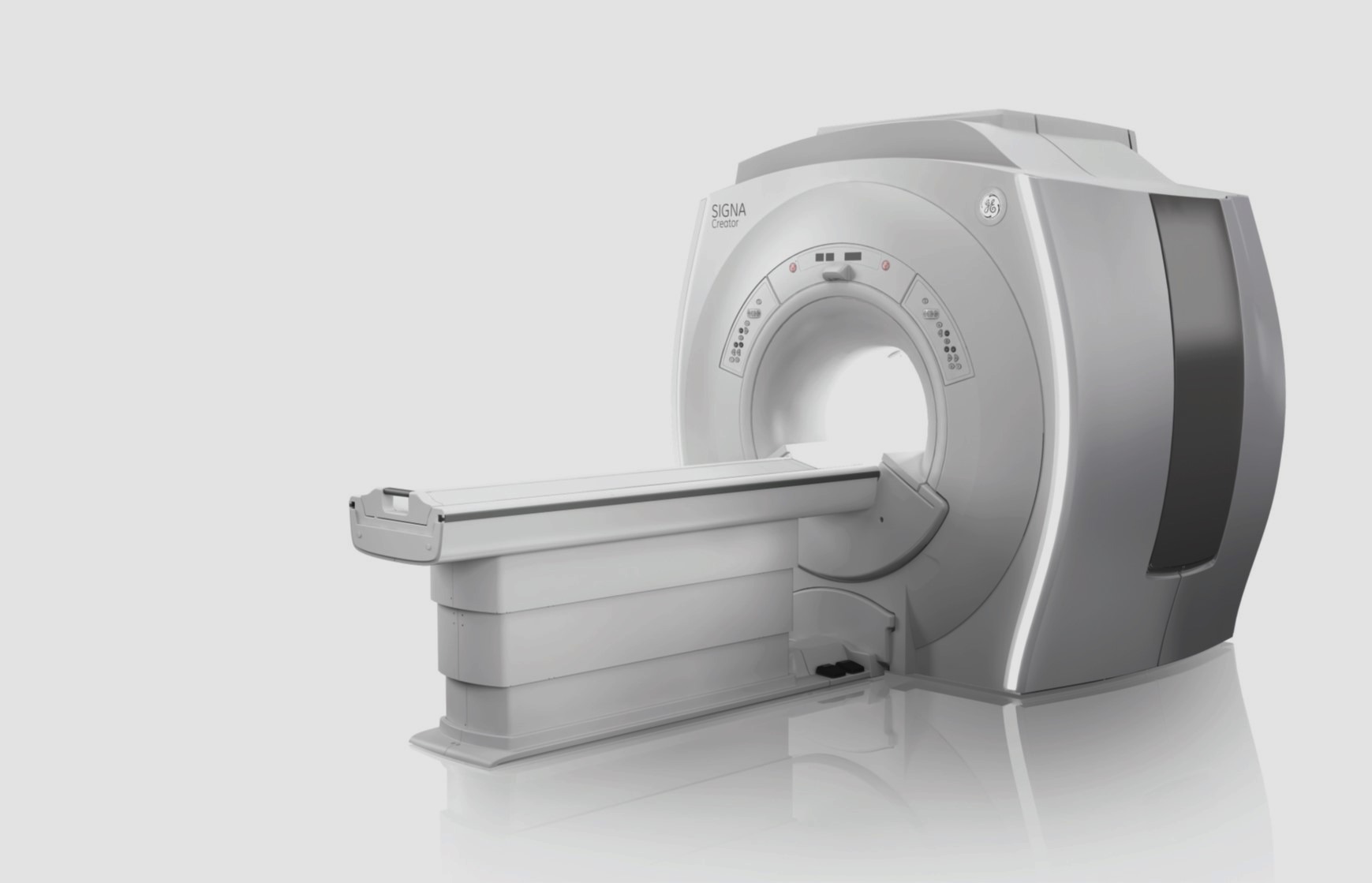
MRI
Magnetic Resonance Imaging (MRI) is a non-radioative imaging examination. The magnetic field produced by MRI forces certain atoms in your body to line up in a particular way. Radio-waves are sent towards these atoms and bounce back, and a computer records the signal produced. Since different types of tissues send back different signals, the computer can render a detailed image of the internal organs. The use of MRI is prominent in the evaluation of neurological status, head & neck, abdomen, pelvis and musculoskeletal conditions. Moreover, it is sensitive in detecting problems in joints, muscles, tendons and ligaments.
Due to the strong magnets used during the imaging, patients must remove any metal from their bodies before getting scanned to avoid accidents. Patients with certain metal implants or fragments such as a cardiac pacemaker, metal spine device, aneurysm clips or hearing aid implants are strongly advised against undergoing the MRI examination.
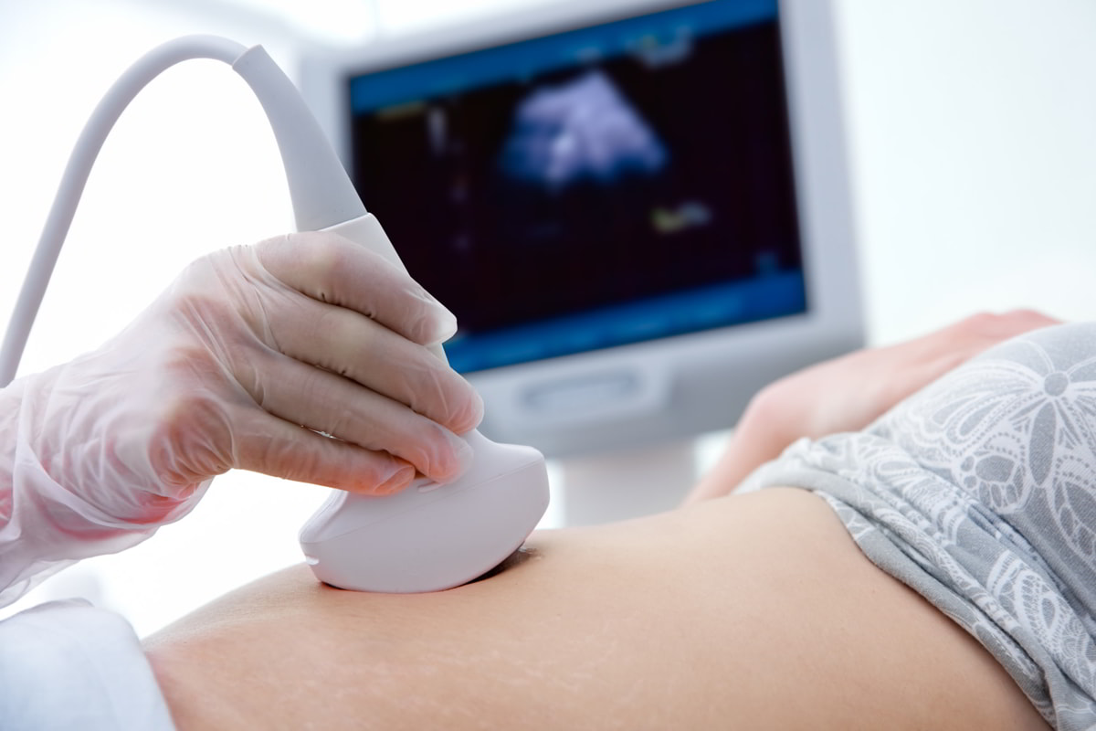
ULTRASOUND
Ultrasound imaging uses high-frequency sound waves which beyond hearing range of human to capture real time pictures of the inside of the body. It involves the use of a small transducer (probe) and ultrasound gel placed directly on the skin. High- frequency sound waves are transmitted from the probe through the gel into the body. The transducer collects the sounds that bounce back and a computer then uses those sound waves to create an image.
Ultrasound image can show the structure and movement of the body's organs, blood flowing through the vessels. Therefore, it has been widely applied to help diagnose diseases of the organs and is also used to help guide biopsies.
Ultrasound is safe, painless, non-radiative and non-invasive. Please consult your physician if an ultrasound imaging is required.

